International Journal of Tropical Medicine
Evaluating the Effects of Spirulina (Arthrospira platensis) on Artemether/Lumefantrine (Coartem)-Induced Oxidative Stress in Wistar Rats
Authors : T.N. Metoh, R.K.A. Siberedi and C.A. Pieme
Abstract: Malaria is considered as a major health problem in Cameroon and other parts of Sub-Saharan Africa. It remains one of the greatest causes of morbidity and mortality in the world with Plasmodium falciparum being the major cause of all deaths. Its management involves the use of conventional drugs with Artemether/Lumefantrine, AL (Coartem) being the firstchoice drug. The administration of coartem not only provides beneficial effects in killing the malaria parasite but also induces oxidative stress which is fatal to the system. Spirulina, a cyanobacteria is used worldwide as a nutraceutical and has potential antioxidant properties and could serve as an adjunct therapy in malaria management. The study was thus designed to evaluate the toxic potential of administration of the therapeutic dose of coartem and the ameliorative effects of spirulina. Rats were treated with distilled water, AL 27 mg/kg and AL 27 mg/kg+ different concentration of spirulina (75, 150 and 300 mg/kg). Oxidative stress was assessed by assessing levels of Malonyldialdehyde (MDA), Total Antioxidant Capacity (TAC), Glutathione (GSH), Alanine Aminotransferase (ALT) and aspartate amino transferase (AST) activities. This study found that AL nor spirulina did not significantly (p<0.05) affect bodyweight. AL caused oxidative stress as indicated by high levels of MDA, ALT and AST activities with a decrease in TAC and GSH levels. The combined treatment of rats with both AL and spirulina showed ameliorative potential of spirulina to oxidative stress induced by AL in a dose dependent manner with spirulina 300 mg/kg showing the greatest protective effects as indicated by a significant (p<0.05) reduced levels of MDA, ALT and AST activities with significant (p<0.05) increases in TAC and GSH levels. These results therefore, suggests that spirulina possesses strong antioxidant capacity and could be used as an adjunct therapy in the management of malaria.
How to cite this article:
T.N. Metoh, R.K.A. Siberedi and C.A. Pieme, 2022. Evaluating the Effects of Spirulina (Arthrospira platensis) on Artemether/Lumefantrine (Coartem)-Induced Oxidative Stress in Wistar Rats. International Journal of Tropical Medicine, 17: 1-9.
INTRODUCTION
Malaria remains one of the leading public health problems in Cameroon as in other parts of Sub-Saharan Africa and Southeastern Asia irrespective of the enhanced management measures[1-3]. Malaria continues to be one of the greatest causes of morbidity and mortality in the world with Plasmodium falciparum being the major cause of all deaths. Globally, there were an estimated 229 million malaria cases in 2019. The World Health Organization (WHO) African Region accounted for about 94% of these cases with 409,000 death cases recorded of which 67% were among children aged under 5 years[3]. The management of malaria involves a collaborative approach which includes preventive measures that aim at controlling the vectors such as use of Insecticide-Treated Nets (ITNs), Indoor Residual Sprays (IRS) and parasite elimination by use of conventional drugs including artemisinin monotherapies and Artemisinin-based Combination Therapies (ACTs)[4-6]. Increasing parasite resistance to ancient malaria drugs such as chloroquine, mefloquine, etc., let to the development and use of ACTs as the recommended drugs of choice used as the first-line of treatment of malaria caused by P. falciparum[7]. Artemether/Lumefantrine (AL) is one of the approved and most successful fixed dose ACT used in the treatment of uncomplicated malaria by P. falciparum. Unfortunately, the partial artemisinin resistance which is characterized by slow parasite clearance during the 3 days treatment with ACTs has resulted to late treatment failures[8]. AL causes malaria parasite death through the generation of free radicals. These excessive generation of free radicals such as Reactive Oxygen Species (ROS) may deplete the antioxidant defense system causing detrimental effects on target cellular components such as DNA, proteins and lipids[9]. Also, there have been recent concerns on their anti-fertility and anti-androgenic effects in experimental animals[10]. The damaging effects of these free radicals are however cushioned by some endogenous antioxidants like glutathione reductase, catalase, superoxide dismutase, etc., these endogenous antioxidants have been found to be insufficient in the complete prevention of the effects of free radicals[11].
In such events, the administration of exogenous antioxidants becomes highly imperative. Unfortunately, many reports have argued that the administration of synthetic antioxidants evoke many side effects[12]. Whereas natural sources of antioxidants exhibit very strong antioxidant effects due to the presence of secondary metabolites, phenols, flavonoids, alkaloids, terpenoids[13]. However, the need to replace synthetic antioxidants with natural ones is on an increase[14]. Spirulina, a cyanobacteria with high antioxidant capacity could therefore, be exploited not only as a food source but as an adjunct therapy to the malaria therapy. Hence, we aimed at evaluating the antioxidant effect of Arthrospira platensis on AL induced-oxidative stress in Wistar rats.
MATERIALS AND METHODS
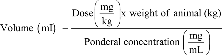
Preparation of spirulina extract: Following the therapeutic dose of spirulina, 10 g/70 kg (142 mg/kg) of body weight, the extract was prepared using the formula below and was then stored in the refrigerator at 4°C:
 |
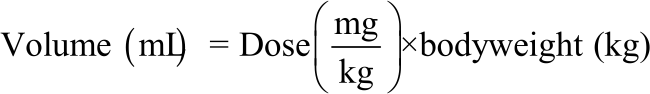
Preparation of AL: Using the therapeutic dose of AL, 27 mg/kg of body weight, the volume to be administered was prepared using the formula below and was then stored in a refrigerator at 4°C:
 |
Preparation/grouping of the rats for the experiment: The 36 healthy wistar rats of three weeks old of mass 100±10 g bought from the Department of Animal Biology of the University of Yaounde I, Cameroon under the Department of Biochemistry, Faculty of Science. These rats were kept in laboratory conditions (12:12 light-dark cycle at approximately 25°C), properly feed and their masses weighed with the aid of an electronic scale balance and recorded. The rats were then grouped into groups of six rats (Table 1).
Among these six groups of rats, the first group designated the “control positive” that took no treatment, the second group designated as “positive control” was administered just AL, the next three “experimental groups” gavaged with an equal volume of AL followed by their respective doses of spirulina extract (75, 150 or 300 mg/kg), the last group was administered spirulina extract at double the therapeutic dose (300 mg/kg).
Treatment of rats: The rats were treated with either distilled water or AL or spirulina extract or a combination of both orally by the use of a gavage tube for 3 days according to the group designated to each. The animals were fasted (i.e., food but not water was withheld for 3 h) prior to administration, the rats were weighed daily until the end of the studies. Animals were observed continuously during the first 30 min after dosing and observed periodically (with special attention given during the first 4 h) for the next 24 h and then daily thereafter.
| Table 1: Parameters of group rate | |||||
| Composition test solution (Groups) | |||||
| 1 | 2 | 3 | 4 | 5 | 6 |
| Distilled water | Therapeutic dose of AL | Therapeutic dose of | Therapeutic dose | Therapeutic dose of AL+ | Double therapeutic dose |
| AL+ half therapeutic | of AL+ therapeutic | double therapeutic dose | of spirulina (300 mg/kg) | ||
| dose of spirulina | dose of spirulina | of spirulina (300 mg/kg) | |||
| (75 mg/kg) | (150 mg/kg) | ||||
Sacrifice and collection of samples: At the end of the 3 days of treatment (on day 4 and 7), the animals were anaesthetized using diazepam (10 mg/mL) at a dose of 10 mg/kg. Blood samples were collected from each rat through the retro-orbital plexus into non-heparinized centrifugal tubes and centrifuged at 3000 rpm for 15 min to obtain the serum fraction. Liver homogenates were also prepared and centrifuged. The serum and liver homogenates were stored in a freezer (-20°C) and were only removed prior to biochemical analysis of the oxidative stress markers.
Malondialdehyde (MDA) concentration: For both serum and liver homogenate, the method used for MDA quantification is that described by Wilbur et al.[15]. Briefly, 100 μL of distilled water (for blank) or sample were placed in a clean test tube followed by the addition of 2000 μL of freshly prepared MDA solution (TCA-TBA-HCL solution) and homogenized. The homogenized mixture was then heated at 100°C for 15 min using a water bath after which it was allowed to cool and then centrifuged at 3000 rpm for 5 minutes before reading the absorbance at 532 nm using a spectrophotometer. The concentration of MDA was determined using its molecular extinction coefficient (ε = 1.56×10⁵ M-1cm-1). The results were determined in μmol/L.
Total Antioxidant Capacity (TAC): TAC was determined by the Ferric Reducing Ability of Plasma (FRAP) assay as previously described[16]. Briefly, 75 μL of distilled water (for blank) or sample was introduced in a clean test tube followed by the addition of 2000 μL of FRAP reagent (acetate buffer, pH 3.6, +tripyridyltriazine solution+FeCl3) and then homogenized. The homogenized mixture was then incubated at ambient temperature for 12 min before reading the absorbance spectrophotometrically at 593 nm. The concentration of the sample was gotten by tracing it back to the FRAP standard curve and the results expressed in mM.
Glutathione concentration: The quantification of glutathione was done following the method as previously described[17]. A volume (20 μL) of sample were introduced into test tubes, followed by 3 mL of Ellman’s reagent (acid-2,2-dithio-5,5'-dibenzoic acid). After homogenization, the tubes were incubated at room temperature for 1 h. In the blank, the samples were replaced by 20 μL of 1M phosphate buffer, pH 7.2. Their ODs read by use of a spectrophotometer at 412 nm against the blank. The amount of cellular glutathione was expressed in μM of protein according to the following formula:
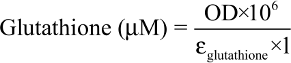 |
Where:
| OD | = | Optical density |
| l | = | Optical path length |
| εglutathione | = | 13600 mole-1.cm-1 |
Alanine aminotransferase or Glutamate Pyruvate
Transaminase (ALT/GPT): Serum ALT activity was performed spectrophotometrically according to the manufacturer’s procedure. Working reagent was prepared by dissolving the contents of R2 substrate (ALT coenzyme. NADH 1.3 mmol/L, 2-oxoglutarate 75 mmol/L. Biocides) in 10,000 μL of R1 buffer (ALT substrate. TRIS buffer 150 mmol/L pH 7.3, L-alanine 750 mmol/L, lactate dehydrogenase>1350 U/L). To begin with 1000 μL of the working reagent was pipetted directly into the cuvette and 100 μL of each serum samples were added. The contents of the cuvettes were mixed and incubated at room temperature for 5 min. The initial absorbance was read, the stopwatch started and the absorbances were read at 1min interval for 3 min. The difference between absorbances and the average absorbances differences per minute were calculated. (All absorbances were read at 340 nm)[18].
Aspartate aminotransferase or Glutamate
Oxaloacetate Transaminase (AST/GOT): Serum AST was performed spectrophotometrically according to the manufacturer’s procedure. Working reagent was prepared by dissolving the contents of R2 substrate (AST coenzyme. NADH 1.3 mmol/L, 2-oxoglutarate 75 mmol/L. Biocides) in 10,000 μL of R1 (AST substrate. TRIS buffer 121 mmol/L pH 7.8, L-aspartate 362 mmol/L, malatedehydrogenase>460 U/L.) buffer. The 1000 μL of the working reagent was pipetted directly into the cuvette and 100 μL of each sample were added. The contents of the cuvettes were mixed and incubated at room temperature for 5 min. The initial absorbance was read, the stopwatch started and the absorbance were read at 1 min interval for 3 min. The difference between absorbances and the average absorbances differences per minute was calculated. (All absorbances were read at 340 nm)[18].
Statistical analysis: Data were expressed as mean± Standard Error of Mean (SEM). They were analysed by one-way analysis of variance (ANOVA) and differences between groups assessed using Student-Newman-Keuls’ test. Differences were considered statistically significant at p<0.05. All analyses were performed using SPSS software package, Version 21.
RESULTS
Effects of AL and spirulina on body weight: Our findings from this study shows that the mean weight of the animals treated with AL varied from 112 g on day 1 and 2, 117 g on day 3, 109 g on day 4-111 g on day 7 with no statistically significant difference when compared to the untreated control group with mean weight ranging from 121 g on day 1-126 g on day 7. Likewise, in the AL+Spirulina 300 mg/kg, the mean weight was 113 g on day 1, 114 g on day 2, 112 g on day 3,111 g on day 4-116 g on day 7 with no statistically significant (p>0.05) difference compared to the control untreated groups (Fig. 1 ).
Effects of combined treatment of AL and Spirulina on
MDA (μM) concentration: The findings from the evaluation of the effects of the combined treatment of AL and spirulina on MDA concentration in both serum and liver homogenate is presented on Table 2. The treatment of the animals with AL 27 mg/kg, AL 27 mg/kg+spirulina 75 mg/kg, AL 27 mg/kg+spirulina 150 mg/kg and AL 27 mg/kg+spirulina 300 mg/kg showed a significant increase (p<0.05) in MDA concentration in serum on day 4 as compared to the normal group while AL 27 mg/kg+spirulina 150 mg/kg, AL 27 mg/kg+ spirulina 300 mg/kg and spirulina 300 mg/kg showed a significant decrease as compared to the group treated with AL 27 mg/kg and on day 7, only groups treated with AL 27 mg/kg, AL 27 mg/kg+ spirulina 75 mg/kg and AL 27 mg/kg+spirulina 150 mg/kg showed a significant increase (p<0.05) with the normal group while a significant decrease was observed within the groups treated with AL 27 mg/kg, AL 27 mg/kg+ spirulina 75 mg/kg and AL 27 mg/kg+spirulina 150 mg/kg for the 2 days. In the liver homogenate, animals treated with AL 27 mg/kg, AL 27 mg/kg+ spirulina 75 mg/kg and AL 27 mg/kg+spirulina 150 mg/kg showed a significant increase (p<0.05) in MDA concentration on day 4 as compared to the normal group while groups treated with AL 27 mg/kg+ spirulina 300 mg/kg and spirulina 300 mg/kg showed a significant decrease as compared to the group treated with AL 27 mg/kg and on day 7, only groups treated with AL 27 mg/kg, AL 27 mg/kg+spirulina 75 mg/kg and AL 27 mg/kg+spirulina 150 mg/kg showed a significant increase (p<0.05) with the normal group while a significant decrease was observed in the groups treated with AL 27 mg/kg+ spirulina 75 mg/kg, AL 27 mg/kg+spirulina 150 mg/kg, AL 27 mg/kg+ spirulina 300 mg/kg and spirulina 300 mg/kg as compared to the group treated with AL 27 mg/kg.
Effects of combined treatment of AL and Spirulina on
TAC (μM) concentration: As presented on Table 3, the findings from this study shows that he treatment of the animals with AL 27 mg/kg+spirulina 150 mg/kg, AL 27 mg/kg+spirulina 300 mg/kg and spirulina 300 mg/kg showed a significant increase (p<0.05) in TAC concentration in serum on day 4 and 7 as compared to the normal group and that treated with AL 27 mg/kg. In the liver homogenate, animals treated with AL 27 mg/kg+spirulina 150 mg/kg, AL 27 mg/kg+spirulina 300 mg/kg and spirulina 300 mg/kg showed a significant increase (p<0.05) in TAC concentration on day 4 as compared to the normal group while only the group treated with spirulina 300 mg/kg showed a significant increase as compared to the group treated with AL 27 mg/kg. Also, there was a significant decrease within the 2 days in each group.
Effects of combined treatment of AL and spirulina on
GSH (μM) concentration: The findings from the evaluation of the effects of the combined treatment of AL and spirulina on GSH in both serum and liver homogenate is presented on Table 4. The treatment of animals with AL 27 mg/kg, AL 27 mg/kg+spirulina 150 mg/kg, AL 27 mg/kg+spirulina 300 mg/kg and spirulina 300 mg/kg increased the concentration of serum GSH as compared to the normal group on day 4 and 7. However, this increase was not statistically significant (p<0.5). In the liver homogenate, animals treated with AL 27 mg/kg+spirulina 150 mg/kg, AL 27 mg/kg+spirulina 300 mg/kg and spirulina 300 mg/kg showed a significant increase (p<0.05) in GSH concentration on day 4 as compared to the normal group and that treated with AL 27 mg/kg. Also, only the group treated with spirulina 300 mg/kg showed a significant decrease within the 2 days in each group.
The effects of combined treatment of AL and spirulina
on serum ALT and AST activity (IU/l): The findings from the evaluation of the effects of the combined treatment of AL and spirulina on ALT and AST in serum is presented on Table 5. Only the treatment of animals with AL 27 mg/kg increased serum ALT activity as compared to the normal group on day 4 while only the group treated with spirulina 300 mg/kg significantly decreased ALT activity as compared to that treated with AL 27 mg/kg. Also, only the groups treated with AL 27 mg/kg and AL 27 mg/kg+ spirulina 75 mg/kg showed a significant decrease within the groups for each day of follow-up.
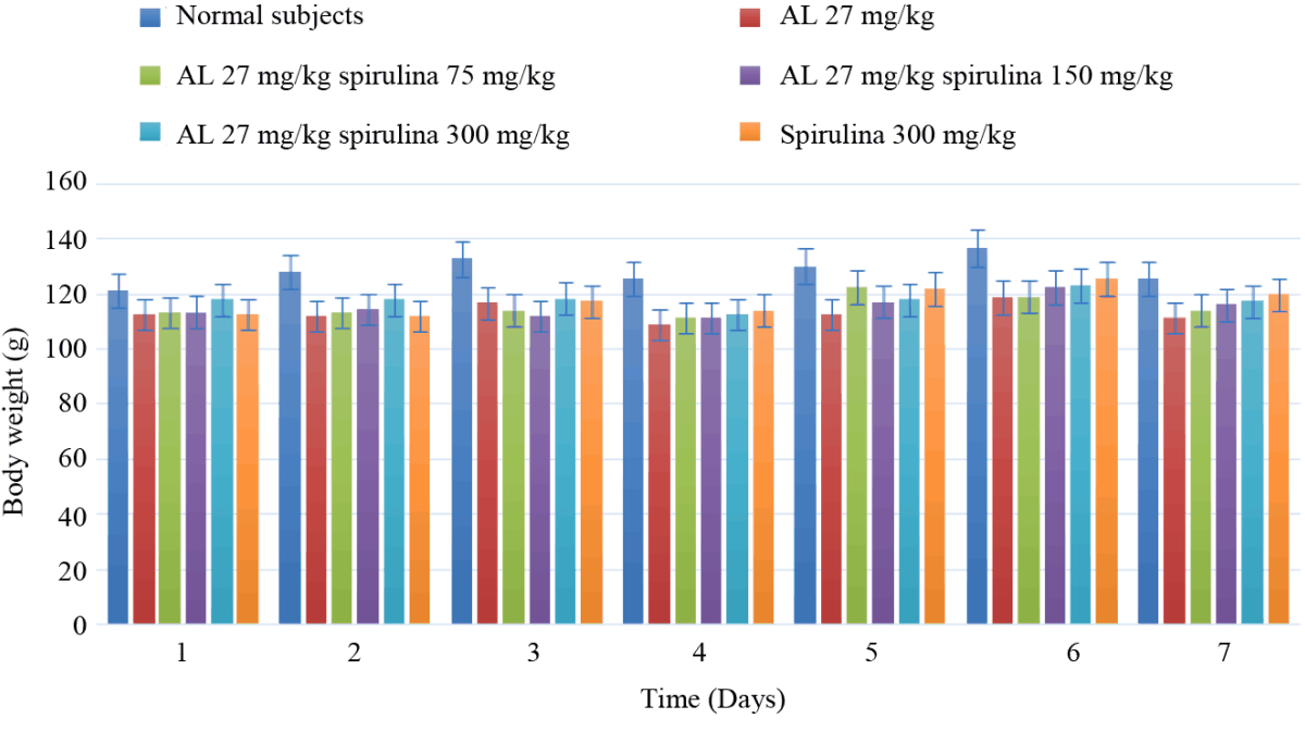 |
| Fig. 1: | Effects of AL and spirulina on body weight |
| Table 2: Effects of combined treatment of AL and spirulina on MDA (μM) concentration | |||||||
| Period | Normal | AL (27 mg/kg)+ | AL (27 mg/kg)+ | AL (27 mg/kg)+ | Spirulina | ||
| Sample | (Days) | subjects | AL (27 mg/kg) | spirulina 75 mg/kg | spirulina 150 mg/kg | spirulina 300 mg/kg | 300 mg/kg |
| Serum | 4 | 0.96±0.07a | 1.50±0.07b | 1.38±0.04bc | 1.34±0.02cd | 1.12±0.05d | 1.02±0.02a |
| 7 | 0.94±0.07a | 1.13±0.09b | 1.18±0.04b | 1.12±0.05bc | 0.98±0.06c | 0.96±0.06a | |
| Liver | 4 | 0.44±0.02a | 0.67±0.04b | 0.59±0.01b | 0.57±0.02b | 0.55±0.04b | 0.40±0.07a |
| 7 | 0.43±0.01a | 0.63±0.05b | 0.53±0.03c | 0.52±0.01c | 0.46±0.04a | 0.44±0.03a | |
| Letters indicate the difference in MDA concentration between the treated groups compared to the normal group at p<0.05 | |||||||
| Table 3: Effects of combined treatment of AL and spirulina on TAC (μM) concentration | |||||||
| Period | Normal | AL (27 mg/kg)+ | AL (27 mg/kg)+ | AL (27 mg/kg)+ | Spirulina | ||
| Sample | (Days) | subjects | AL (27 mg/kg) | spirulina 75 mg/kg | spirulina 150 mg/kg | spirulina 300 mg/kg | 871.55±21.43c |
| Serum | 4 | 529.77±23.05a | 543.77±13.47a | 572.66±20.81a | 611.55±18.35b | 801.55±9.62bc | 829.33±30.55c |
| 7 | 586.00±25.16a | 640.44±19.24a | 632.66±15.27a | 719.22±27.46b | 772.00±36.71bc | 795.99±33.33b | |
| Liver | 4 | 632.66±28.48a | 686.33±23.83a | 686.00±12.01a | 698.44±19.32a | 728.66±30.78ab | 0.40±0.07a |
| 7 | 520.33±15.58a | 541.66±21.36a | 527.11±36.71ca | 544.66±51.73a | 567.33±38.33a | 615.66±41.63a | |
| Letters indicate the difference in TAC concentration between the treated groups compared to the normal group at p<0.05 | |||||||
| Table 4: Effects of combined treatment of AL and spirulina on GSH (μM) concentration | |||||||
| Period | Normal | AL (27 mg/kg)+ | AL (27 mg/kg)+ | AL (27 mg/kg)+ | Spirulina | ||
| Sample | (Days) | subjects | AL (27 mg/kg) | Spirulina 75 mg/kg | Spirulina 150 mg/kg | Spirulina 300 mg/kg | 300 mg/kg |
| Serum | 4 | 2.45±0.42a | 3.18±0.42a | 3.43±0.42a | 3.43±0.42a | 3.43±0.42a | 2.69±0.42a |
| 7 | 2.45±0.42a | 2.94±0.73a | 3.18±0.42a | 2.94±0.73a | 2.69±0.42a | 2.69±0.42a | |
| Liver | 4 | 2.57±0.36a | 2.69±0.42a | 3.18±0.42ab | 3.67±0.00b | 3.92±0.42b | 3.67±0.00b |
| 7 | 2.69±0.42a | 2.94±0.73a | 3.18±0.42a | 3.67±0.73a | 4.16±0.42a | 3.30±0.36ca | |
| Letters indicate the difference in GSH concentration between the treated groups compared to the normal group at p<0.05 | |||||||
| Table 5: The effects of combined treatment of AL and spirulina on serum ALT and AST activity (IU/l) | |||||||
| Period | Normal | AL (27 mg/kg)+ | AL (27 mg/kg)+ | AL (27 mg/kg)+ | Spirulina | ||
| Enzyme | (Days) | subjects | AL (27 mg/kg) | Spirulina 75 mg/kg | Spirulina 150 mg/kg | Spirulina 300 mg/kg | 300 mg/kg |
| ALT | 4 | 119.07±8.77a | 142.99±3.82b | 139.91±7.35b | 130.14±8.77b | 120.86±13.75a | 121.96±9.07a |
| 7 | 123.38±8.61a | 134.26±6.65a | 129.61±0.70a | 126.29±11.11a | 125.89±16.12a | 125.88±8.46a | |
| AST | 4 | 555.98±59.17a | 820.79±67.13b | 703.40±35.42b | 638.97±82.28a | 633.21±78.98a | 572.68±17.46a |
| 7 | 574.60±48.68a | 690.07±20.46a | 678.26±20.46a | 659.28±38.03a | 634.78±74.12a | 576.23±45.76a | |
| Letters indicate the difference in ALT and AST concentration between the treated groups compared to the normal group at p<0.05 | |||||||
On day 4, AST activity increased significantly (p<0.05) in the groups treated with AL 27 mg/kg and AL 27 mg/kg+spirulina 75 mg/kg treated group. The reverse was observed with a significant (p<0.05) decrease of AST activity in the groups treated either with AL 27 mg/kg+spirulina 150 mg/kg or AL 27 mg/kg+ spirulina 300 mg/kg or spirulina 300 mg/kg as compared to that of the AL 27mg/kg treated group.
DISCUSSION
The p resent study has shown that, the administration of AL and/or spirulina does not significantly affect body weight. This may be due to spirulina’shypolipidemic and hypo cholesterol properties that prevented the animals from gaining significant weight consistent with the findings of previous studies[19-21]. Also, the insignificant change in body weight in the group treated with AL alone and the insignificant difference between day 4 and day 7 weights per group further supports the fact that spirulina doesn’t affect body weight within the treatment days.
Administration of AL and spirulina affects oxidative stress status of animals as observed by evaluating oxidative stress markers. In this study, the administration of distilled water was to obtain a base line of oxidative stress status whereas the administration of AL was to induce oxidative stress. This then gave the room to evaluate the protective effects of spirulina on the animals where they were treated with both AL and different concentrations of spirulina under the same conditions for a period of 3 days, following the 3 days regimen for the therapeutic treatment of malaria.
Malondialdehyde (MDA) is an organic compound (colorless liquid) with the nominalformula CH2(CHO)2, it is a highly reactive compound that occurs as an enol and formed as one of the final products of polyunsaturated fatty acids peroxidation in the cells. It occurs naturally and is a marker for oxidative stress[22]. From MDA results, AL administration had a significant increase in MDA concentration on both day 4 and 7 as compared to distilled water in both serum and liver, illustrating the potential oxidative stress caused by AL. These results were in line with previous studies[23-27]. Thus, signifying a high level of lipid peroxidation induced by AL. However, there was a significant decrease in MDA concentration in animals treated with AL 27 mg/kg+ spirulina 150 mg/kg, AL 27 mg/kg+ spirulina 300 mg/kg and spirulina 300 mg/kg as compared with those treated with AL 27 mg/kg for day 4. These results indicate that spirulina decreases (dose dependent) lipid peroxidation which has also been reported by previous studies[28-30]. The decrease in MDA concentration within the groups on day 4 and 7 is because administration of AL was stopped on day 3 of the study.
Ferric Reducing Antioxidant Power Assay (FRAP), the assay measures the antioxidant potential in samples through the reduction of ferric iron (Fe3+) to ferrous iron (Fe2+) by antioxidants present in the samples. The greater the concentration, the greater the antioxidants present in the sample[31]. From FRAP results, there was an increase in serum and liver TAC in the group treated with AL 27 mg/kg as compared to the normal subjects though not significant, this could be due to the fact that the system’s antioxidant molecules are being deployed to counteract the free radicals produced by AL. There was an increase in TAC in the groups treated with 27 mg/kg+ spirulina 75 mg/kg, 27 mg/kg+ spirulina 150 mg/kg, AL 27 mg/kg+ spirulina 300 mg/kg and spirulina 300 mg/kg as compared to the animals treated with AL 27 mg/kg. However, this increase was only significant in the groups treated with 27 mg/kg+ spirulina 150 mg/kg, AL 27 mg/kg+ spirulina 300mg/kg and spirulina 300 mg/kg and was dose dependent. These results signify that spirulina has a great antioxidant capacity[29, 32-35] and this could be related to the presence of molecules such as polyphenols, polysaccharides, flavonoids, phycocyanin and vitamins that greatly contribute to its antioxidant property[36-37].
Glutathione is produced by the liver and involved in many body processes like tissue building and repair, making chemicals and proteins needed in the body and in immune system function. It is asubstance made from the amino acids glycine, cysteine and glutamic acid. Glutathione exists in cells in 2 states: reduced (GSH) and oxidized (GSSG) the ratio of GSH to GSSG determines cell redox status of cells. Healthy cells at rest have a GSH/GSSG ratio >100 while the ratio drops to 1-10 in cells exposed to oxidant stress, it plays a crucial role in shielding cellular macromolecules from endogenous and exogenous reactive oxygen and nitrogen species[38]. Results from this study shows that there was an increase in serum GSH concentration on day 4 in the group treated with AL 27 mg/kg as compared with the normal subjects though not significant. The increase could be explained by the fact that AL induced GSH production in response to the oxidative stress it induced. Also, there was an increase in the rest of the groups as compared to the normal and AL 27 mg/kg groups which was dose dependent. This increase could be due to response to oxidative stress induced by AL and the administration of spirulina which is a very rich source of GSH, also the insignificant increase observed in these groups could be due to the short period of treatment in the animals. In the liver, an insignificant increase in GSH concentration was also observed in the groups treated with AL 27 mg/kg and AL 27 mg/kg+ spirulina 75 mg/kg whereas a significant increase was observed in the groups treated with 27 mg/kg+ spirulina 150 mg/kg, AL 27 mg/kg+ spirulina 300 mg/kg and spirulina 300 mg/kg in a dose dependent manner. The increase in these groups was also significant as compared to the group treated with just AL 27 mg/mg and is explained by the fact that spirulina is a very rich source of GSH[38]. These increment in GSH concentration were in conformity with those of previous studies[39-41]. The increase in GSH in the liver as compared to serum can be backed by the fact that GSH is being produced in the liver and is concentrated more in the liver than in blood[38].
Alanine aminotransferase, usually referred to as ALT and Aspartate aminotransferase (AST), are enzymes that are concentrated primarily in the liver[18]. An ALT or AST test measures the amount of these enzymes in the blood. ALT and AST levels can increase when liver cells are damaged, so, the tests can be used to evaluate the condition of the liver[42-44]. Results from the present study shows that there was a significant increase in ALT and AST activity in the group treated with AL 27 mg/kg (and AL 27 mg/kg+ spirulina 75 mg/kg for group for AST) as compared to the normal subjects whereas there was a decrease in ALT activity observed in the AL 27 mg/kg+ spirulina 75 mg/kg and AL 27 mg/kg+ spirulina 150 mg/kg groups though not significant. However, there was a significant decrease in ALT activity in the AL 27 mg/kg+ spirulina 300 mg/kg and spirulina 300 mg/kg treated groups and AL 27 mg/kg+ spirulina 150 mg/kg, AL 27 mg/kg+ spirulina 300 mg/kg and spirulina 300 mg/kg treated groups for AST as compared to the AL 27 mg/kg group. This decrease was dose dependent. These results were in conformity with previous findings[45-48] which revealed that AL is capable of inducing liver damage with high levels of ALT and AST due to production of ROS. Interesting, the results of this study are consistent with previous findings[39, 49] that indicated that administration of spirulina is capable of decreasing ALT and AST activities meaning that Arthrospira platensis could be involved in the prevention of liver damage.
CONCLUSION
The therapeutic dose of AL increased MDA concentration, ALT and AST activities in both serum and liver thus indicating potential oxidative stress. Arthrospira platensis increased glutathione concentration and total antioxidant capacities as well as reduced malondialdehyde, ALT and AST activity in the extract-treated groups induced with AL, demonstrating that Arthrospira platensis has protective effect against oxidative stress. Hence, Arthrospira platensis could be used not just as a food but also as an adjunct therapy in the medical domain to curb oxidative stress caused by AL used in the management of malaria or other oxidative stress inducers.
Abbreviations:
| • | ACT: Artemisinin-based combinational therapy |
| • | AL: Artemether/Lumefantrine |
| • | ALT: Alanine aminotransferase |
| • | AST: Aspartate aminotransferase |
| • | FDA: Food and Drug Administration |
| • | FRAP: Ferric reducing ability of plasma |
| • | GSH: Glutathione |
| • | MDA: Malonyldialdehyde |
| • | OD: Optical density |
| • | PCR: Polymerase chain reaction |
| • | RDT: Rapid diagnostic test |
| • | RNS: Reactive nitrogen species |
| • | ROS: Reactive oxygen species |
| • | TAC: Total antioxidant capacity |
| • | WHO: World Health Organization |
ACKNOWLEDGMENTS
The authors are grateful to the Dean of the Faculty of Medicine and Biochemical Sciences (FMBS) of the University of Yaounde I for providing the necessary platform to perform this research work.
Authors’ contributions: MTN conceived and designed the study. MTN and PCA coordinated the study. SR performed the Biochemical analysis and data collection. MTN prepared and review the manuscript. PCA reviewed the manuscript. MTN critically revised the manuscript. All authors read and approved the final manuscript.
Ethics approval and consent to participate: Not applicable.
Competing interests: The authors declare that they have no competing interests.